General Information About Skin Cancer
Skin cancer is a disease in which malignant (cancer) cells form in the tissues of the skin.
The skin is the body's largest organ. It protects against heat, sunlight, injury, and infection. Skin also helps control body temperature and stores water, fat, and vitamin D. The skin has several layers, but the two main layers are the epidermis (upper or outer layer) and the dermis (lower or inner layer). Skin cancer begins in the epidermis, which is made up of three kinds of cells:
- Squamous cells: Thin, flat cells that form the top layer of the epidermis.
- Basal cells: Round cells under the squamous cells.
- Melanocytes: Cells that make melanin and are found in the lower part of the epidermis. Melanin is the pigment that gives skin its natural color. When skin is exposed to the sun, melanocytes make more pigment and cause the skin to darken.
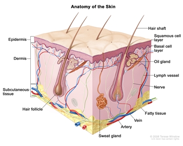
Anatomy of the skin showing the epidermis (including the squamous cell and basal cell layers), dermis, subcutaneous tissue, and other parts of the skin.
Skin cancer can occur anywhere on the body, but it is most common in skin that is often exposed to sunlight, such as the face, neck, hands, and arms.
There are different types of cancer that start in the skin.
The most common types are basal cell carcinoma and squamous cell carcinoma, which are nonmelanoma skin cancers. Nonmelanoma skin cancers rarely spread to other parts of the body. Melanoma is a much rarer type of skin cancer. It is more likely to invade nearby tissues and spread to other parts of the body. Actinic keratosis is a skin condition that sometimes becomes squamous cell carcinoma.
This summary is about nonmelanoma skin cancer and actinic keratosis. See the following PDQ summaries for information on melanoma and other kinds of cancer that affect the skin:
- Melanoma Treatment
- Mycosis Fungoides and the Sézary Syndrome Treatment
- Kaposi Sarcoma Treatment
- Merkel Cell Carcinoma Treatment
- Unusual Cancers of Childhood Treatment
- Genetics of Skin Cancer
Skin color and being exposed to sunlight can increase the risk of nonmelanoma skin cancer and actinic keratosis.
Anything that increases your chance of getting a disease is called a risk factor. Having a risk factor does not mean that you will get cancer; not having risk factors doesn't mean that you will not get cancer. Talk with your doctor if you think you may be at risk. Risk factors for basal cell carcinoma and squamous cell carcinoma include the following:
- Being exposed to natural sunlight or artificial sunlight (such as from tanning beds) over long periods of time.
- Having a fair complexion, which includes the following:
- Fair skin that freckles and burns easily, does not tan, or tans poorly.
- Blue or green or other light-colored eyes.
- Red or blond hair.
- Having actinic keratosis.
- Past treatment with radiation.
- Having a weakened immune system.
- Having certain changes in the genes that are linked to skin cancer.
- Being exposed to arsenic.
Risk factors for actinic keratosis include the following:
- Being exposed to natural sunlight or artificial sunlight (such as from tanning beds) over long periods of time.
- Having a fair complexion, which includes the following:
- Fair skin that freckles and burns easily, does not tan, or tans poorly.
- Blue or green or other light-colored eyes.
- Red or blond hair.
Nonmelanoma skin cancer and actinic keratosis often appear as a change in the skin.
Not all changes in the skin are a sign of nonmelanoma skin cancer or actinic keratosis. Check with your doctor if you notice any changes in your skin.
Signs of nonmelanoma skin cancer include the following:
- A sore that does not heal.
- Areas of the skin that are:
- Raised, smooth, shiny, and look pearly.
- Firm and look like a scar, and may be white, yellow, or waxy.
- Raised, and red or reddish-brown.
- Scaly, bleeding or crusty.
Signs of actinic keratosis include the following:
- A rough, red, pink, or brown, raised, scaly patch on the skin that may be flat or raised.
- Cracking or peeling of the lower lip that is not helped by lip balm or petroleum jelly.
Tests or procedures that examine the skin are used to detect (find) and diagnose nonmelanoma skin cancer and actinic keratosis.
The following procedures may be used:
- Skin exam: A doctor or nurse checks the skin for bumps or spots that look abnormal in color, size, shape, or texture.
- Skin biopsy: All or part of the abnormal-looking growth is cut from the skin and viewed under a microscope by a pathologist to check for signs of cancer. There are four main types of skin biopsies:
- Shave biopsy: A sterile razor blade is used to "shave-off" the abnormal-looking growth.
- Punch biopsy: A special instrument called a punch or a trephine is used to remove a circle of tissue from the abnormal-looking growth.
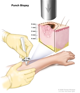
Punch biopsy. A hollow, circular scalpel is used to cut into a lesion on the skin. The instrument is turned clockwise and counterclockwise to cut down about 4 millimeters (mm) to the layer of fatty tissue below the dermis. A small sample of tissue is removed to be checked under a microscope. Skin thickness is different on different parts of the body. - Incisional biopsy: A scalpel is used to remove part of a growth.
- Excisional biopsy: A scalpel is used to remove the entire growth.
Certain factors affect prognosis (chance of recovery) and treatment options.
The prognosis (chance of recovery) depends mostly on the stage of the cancer and the type of treatment used to remove the cancer.
Treatment options depend on the following:
- The stage of the cancer (whether it has spread deeper into the skin or to other places in the body).
- The type of cancer.
- The size of the tumor and what part of the body it affects.
- The patient's general health.
Stages of Skin Cancer
After nonmelanoma skin cancer has been diagnosed, tests are done to find out if cancer cells have spread within the skin or to other parts of the body.
The process used to find out if cancer has spread within the skin or to other parts of the body is called staging. The information gathered from the staging process determines the stage of the disease. It is important to know the stage in order to plan treatment.
The following tests and procedures may be used in the staging process:
- CT scan (CAT scan): A procedure that makes a series of detailed pictures of areas inside the body, taken from different angles. The pictures are made by a computer linked to an x-ray machine. A dye may be injected into a vein or swallowed to help the organs or tissues show up more clearly. This procedure is also called computed tomography, computerized tomography, or computerized axial tomography.
- MRI (magnetic resonance imaging): A procedure that uses a magnet, radio waves, and a computer to make a series of detailed pictures of areas inside the body. This procedure is also called nuclear magnetic resonance imaging (NMRI).
- Lymph node biopsy: For squamous cell carcinoma, the lymph nodes may be removed and checked to see if cancer has spread to them.
There are three ways that cancer spreads in the body.
Cancer can spread through tissue, the lymph system, and the blood:
- Tissue. The cancer spreads from where it began by growing into nearby areas.
- Lymph system. The cancer spreads from where it began by getting into the lymph system. The cancer travels through the lymph vessels to other parts of the body.
- Blood. The cancer spreads from where it began by getting into the blood. The cancer travels through the blood vessels to other parts of the body.
Cancer may spread from where it began to other parts of the body.
When cancer spreads to another part of the body, it is called metastasis. Cancer cells break away from where they began (the primary tumor) and travel through the lymph system or blood.
- Lymph system. The cancer gets into the lymph system, travels through the lymph vessels, and forms a tumor (metastatic tumor) in another part of the body.
- Blood. The cancer gets into the blood, travels through the blood vessels, and forms a tumor (metastatic tumor) in another part of the body.
The metastatic tumor is the same type of cancer as the primary tumor. For example, if skin cancer spreads to the lung, the cancer cells in the lung are actually skin cancer cells. The disease is metastatic skin cancer, not lung cancer.
Staging of nonmelanoma skin cancer depends on whether the tumor has certain "high-risk" features and if the tumor is on the eyelid.
Staging for nonmelanoma skin cancer that is on the eyelid is different from staging for nonmelanoma skin cancer that affects other parts of the body.
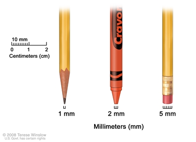
Millimeters (mm). A sharp pencil point is about 1 mm, a new crayon point is about 2 mm, and a new pencil eraser is about 5 mm.
The following are high-risk features for nonmelanoma skin cancer that is not on the eyelid:
- The tumor is thicker than 2 millimeters.
- The tumor is described as Clark level IV (has spread into the lower layer of the dermis) or Clark level V (has spread into the layer of fat below the skin).
- The tumor has grown and spread along nerve pathways.
- The tumor began on an ear or on a lip that has hair on it.
- The tumor has cells that look very different from normal cells under a microscope.
The following stages are used for nonmelanoma skin cancer that is not on the eyelid:
Stage 0 (Carcinoma in Situ)

Stage 0 nonmelanoma skin carcinoma in situ. Abnormal cells are shown in the epidermis (outer layer of the skin).
In stage 0, abnormal cells are found in the squamous cell or basal cell layer of the epidermis (topmost layer of the skin). These abnormal cells may become cancer and spread into nearby normal tissue. Stage 0 is also called carcinoma in situ.
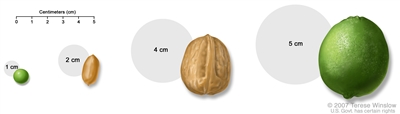
Pea, peanut, walnut, and lime show tumor sizes.
Stage I
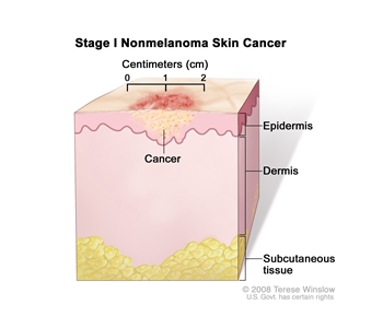
Stage I nonmelanoma skin cancer. The tumor is no more than 2 centimeters.
In stage I, cancer has formed. The tumor is not larger than 2 centimeters at its widest point and may have one high-risk feature.
Stage II
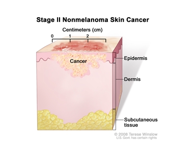
Stage II nonmelanoma skin cancer. The tumor is more than 2 centimeters wide.
In stage II, the tumor is either:
- larger than 2 centimeters at its widest point; or
- any size and has two or more high-risk features.
Stage III
In stage III:
- The tumor has spread to the jaw, eye socket, or side of the skull. Cancer may have spread to one lymph node on the same side of the body as the tumor. The lymph node is not larger than 3 centimeters.
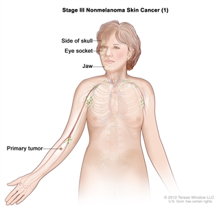
Stage III nonmelanoma skin cancer (1). Cancer has spread from the primary tumor to bones of the jaw, eye socket, or side of the skull.
or
- Cancer has spread to one lymph node on the same side of the body as the tumor. The lymph node is not larger than 3 centimeters and one of the following is true:
- the tumor is not larger than 2 centimeters at its widest point and may have one high-risk feature; or
- the tumor is larger than 2 centimeters at its widest point; or
- the tumor is any size and has two or more high-risk features.
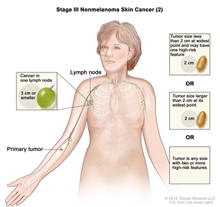
Stage III nonmelanoma skin cancer (2). Cancer has spread to one lymph node that is 3 centimeters or smaller and is on the same side of the body as the primary tumor. Also, the tumor is 2 centimeters or smaller at its widest point and may have one high-risk feature; OR the tumor is larger than 2 centimeters at its widest point; OR the tumor is any size and has two or more high-risk features. There are five high-risk features: (1) the tumor is thicker than 2 millimeters; (2) the tumor has spread into the lower layer of the skin or into the layer of fat below the skin; (3) the tumor has grown and spread along nerve pathways; (4) the tumor began on an ear or on a lip that has hair on it; and (5) the tumor has cells that look very different from normal cells under a microscope.
Stage IV
In stage IV, one of the following is true:
- The tumor is any size and may have spread to the jaw, eye socket, or side of the skull. Cancer has spread to one lymph node on the same side of the body as the tumor and the affected node is larger than 3 centimeters but not larger than 6 centimeters, or cancer has spread to more than one lymph node on one or both sides of the body and the affected nodes are not larger than 6 centimeters; or
- The tumor is any size and may have spread to the jaw, eye socket, skull, spine, or ribs. Cancer has spread to one lymph node that is larger than 6 centimeters; or
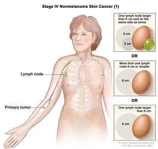
Stage IV nonmelanoma skin cancer (1). The tumor is any size. Cancer has spread to one lymph node that is larger than 3 centimeters but not larger than 6 centimeters and is on the same side of the body as the tumor; OR to more than one lymph node 6 centimeters or smaller on one or both sides of the body; OR to one lymph node that is larger than 6 centimeters. - The tumor is any size and has spread to the base of the skull, spine, or ribs. Cancer may have spread to the lymph nodes; or
- Cancer has spread to other parts of the body, such as the lung.
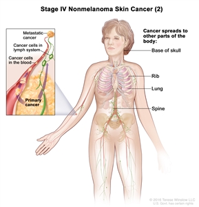
Stage IV nonmelanoma skin cancer (2). The tumor is any size and has spread to the base of the skull, spine, ribs, lung, or other parts of the body.
The following stages are used for nonmelanoma skin cancer on the eyelid:
Stage 0 (Carcinoma in Situ)
In stage 0, abnormal cells are found in the epidermis (topmost layer of the skin). These abnormal cells may become cancer and spread into nearby normal tissue. Stage 0 is also called carcinoma in situ.
Stage I
Stage I is divided into stages IA, IB, and IC.
- Stage IA: The tumor is 5 millimeters or smaller and has not spread to the connective tissue of the eyelid or to the edge of the eyelid where the lashes are.
- Stage IB: The tumor is larger than 5 millimeters but not larger than 10 millimeters or has spread to the connective tissue of the eyelid, or to the edge of the eyelid where the lashes are.
- Stage IC: The tumor is larger than 10 millimeters but not larger than 20 millimeters or has spread through the full thickness of the eyelid.
Stage II
In stage II, one of the following is true:
- The tumor is larger than 20 millimeters.
- The tumor has spread to nearby parts of the eye or eye socket.
- The tumor has spread to spaces around the nerves in the eyelid.
Stage III
Stage III is divided into stages IIIA, IIIB, and IIIC.
- Stage IIIA: To remove all of the tumor, the whole eye and part of the optic nerve must be removed. The bone, muscles, fat, and connective tissue around the eye may also be removed.
- Stage IIIB: The tumor may be anywhere in or near the eye and has spread to nearby lymph nodes.
- Stage IIIC: The tumor has spread to structures around the eye or in the face, or to the brain, and cannot be removed in surgery.
Stage IV
The tumor has spread to distant parts of the body.
Treatment is based on the type of nonmelanoma skin cancer or other skin condition diagnosed:
Basal cell carcinoma
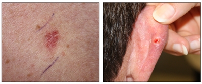
Basal cell carcinoma. A skin cancer lesion that looks reddish brown and slightly raised (left panel) and a skin cancer lesion that looks like an open sore with a pearly rim (right panel).
Basal cell carcinoma is the most common type of skin cancer. It usually occurs on areas of the skin that have been in the sun, most often the nose. Often this cancer appears as a raised bump that looks smooth and pearly. Another type looks like a scar and is flat and firm and may be white, yellow, or waxy. Basal cell carcinoma may spread to tissues around the cancer, but it usually does not spread to other parts of the body.
Squamous cell carcinoma
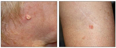
Squamous cell carcinoma. A skin cancer lesion on the face that looks raised and crusty (left panel) and a skin cancer lesion on the leg that looks pink and raised (right panel).
Squamous cell carcinoma occurs on areas of the skin that have been in the sun, such as the ears, lower lip, and the back of the hands. Squamous cell carcinoma may also appear on areas of the skin that have been burned or exposed to chemicals or radiation. Often this cancer appears as a firm red bump. The tumor may feel scaly, bleed, or form a crust. Squamous cell tumors may spread to nearby lymph nodes. Squamous cell carcinoma that has not spread can usually be cured.
Actinic keratosis
Actinic keratosis is a skin condition that is not cancer, but sometimes changes into squamous cell carcinoma. It usually occurs in areas that have been exposed to the sun, such as the face, the back of the hands, and the lower lip. It looks like rough, red, pink, or brown scaly patches on the skin that may be flat or raised, or the lower lip cracks and peels and is not helped by lip balm or petroleum jelly.
Treatment Option Overview
There are different types of treatment for patients with nonmelanoma skin cancer and actinic keratosis.
Different types of treatment are available for patients with nonmelanoma skin cancer and actinic keratosis. Some treatments are standard (the currently used treatment), and some are being tested in clinical trials. A treatment clinical trial is a research study meant to help improve current treatments or obtain information on new treatments for patients with cancer. When clinical trials show that a new treatment is better than the standard treatment, the new treatment may become the standard treatment. Patients may want to think about taking part in a clinical trial. Some clinical trials are open only to patients who have not started treatment.
Six types of standard treatment are used:
Surgery
One or more of the following surgical procedures may be used to treat nonmelanoma skin cancer or actinic keratosis:
- Mohs micrographic surgery: The tumor is cut from the skin in thin layers. During surgery, the edges of the tumor and each layer of tumor removed are viewed through a microscope to check for cancer cells. Layers continue to be removed until no more cancer cells are seen. This type of surgery removes as little normal tissue as possible and is often used to remove skin cancer on the face.
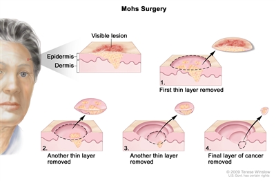
Mohs surgery. A surgical procedure to remove skin cancer in several steps. First, a thin layer of cancerous tissue is removed. Then, a second thin layer of tissue is removed and viewed under a microscope to check for cancer cells. More layers are removed one at a time until the tissue viewed under a microscope shows no remaining cancer. This type of surgery is used to remove as little normal tissue as possible and is often used to remove skin cancer on the face. - Simple excision: The tumor is cut from the skin along with some of the normal skin around it.
- Shave excision: The abnormal area is shaved off the surface of the skin with a small blade.
- Electrodesiccation and curettage: The tumor is cut from the skin with a curette (a sharp, spoon-shaped tool). A needle-shaped electrode is then used to treat the area with an electric current that stops the bleeding and destroys cancer cells that remain around the edge of the wound. The process may be repeated one to three times during the surgery to remove all of the cancer.
- Cryosurgery: A treatment that uses an instrument to freeze and destroy abnormal tissue, such as carcinoma in situ. This type of treatment is also called cryotherapy.
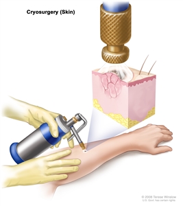
Cryosurgery. An instrument with a nozzle is used to spray liquid nitrogen or liquid carbon dioxide to freeze and destroy abnormal tissue. - Laser surgery: A surgical procedure that uses a laser beam (a narrow beam of intense light) as a knife to make bloodless cuts in tissue or to remove a surface lesion such as a tumor.
- Dermabrasion: Removal of the top layer of skin using a rotating wheel or small particles to rub away skin cells.
Radiation therapy
Radiation therapy is a cancer treatment that uses high-energy x-rays or other types of radiation to kill cancer cells or keep them from growing. There are two types of radiation therapy:
- External radiation therapy uses a machine outside the body to send radiation toward the cancer.
- Internal radiation therapy uses a radioactive substance sealed in needles, seeds, wires, or catheters that are placed directly into or near the cancer.
The way the radiation therapy is given depends on the type of cancer being treated. External radiation therapy is used to treat skin cancer.
Chemotherapy
Chemotherapy is a cancer treatment that uses drugs to stop the growth of cancer cells, either by killing the cells or by stopping them from dividing. When chemotherapy is taken by mouth or injected into a vein or muscle, the drugs enter the bloodstream and can reach cancer cells throughout the body (systemic chemotherapy). When chemotherapy is placed directly into the cerebrospinal fluid, an organ, or a body cavity such as the abdomen, the drugs mainly affect cancer cells in those areas (regional chemotherapy). Chemotherapy for nonmelanoma skin cancer and actinic keratosis is usually topical (applied to the skin in a cream or lotion). The way the chemotherapy is given depends on the condition being treated.
Retinoids (drugs related to vitamin A) are sometimes used to treat squamous cell carcinoma of the skin.
See Drugs Approved for Basal Cell Carcinoma for more information.
Photodynamic therapy
Photodynamic therapy (PDT) is a cancer treatment that uses a drug and a certain type of laser light to kill cancer cells. A drug that is not active until it is exposed to light is injected into a vein. The drug collects more in cancer cells than in normal cells. For skin cancer, laser light is shined onto the skin and the drug becomes active and kills the cancer cells. Photodynamic therapy causes little damage to healthy tissue.
Biologic therapy
Biologic therapy is a treatment that uses the patient's immune system to fight cancer. Substances made by the body or made in a laboratory are used to boost, direct, or restore the body's natural defenses against cancer. This type of cancer treatment is also called biotherapy or immunotherapy.
Interferon and imiquimod are biologic agents used to treat skin cancer. Interferon (by injection) may be used to treat squamous cell carcinoma of the skin. Topical imiquimod therapy (a cream applied to the skin) may be used to treat some small basal cell carcinomas.
See Drugs Approved for Basal Cell Carcinoma for more information.
Targeted therapy
Targeted therapy is a type of treatment that uses drugs or other substances to attack cancer cells. Targeted therapies usually cause less harm to normal cells than chemotherapy or radiation therapy do.
Targeted therapy with a signal transduction inhibitor is used to treat basal cell carcinoma. Signal transduction inhibitors block signals that are passed from one molecule to another inside a cell. Blocking these signals may kill cancer cells. Vismodegib and sonidegib are signal transduction inhibitors used to treat basal cell carcinoma.
See Drugs Approved for Basal Cell Carcinoma for more information.
New types of treatment are being tested in clinical trials.
Information about clinical trials is available from the NCI website.
Patients may want to think about taking part in a clinical trial.
For some patients, taking part in a clinical trial may be the best treatment choice. Clinical trials are part of the cancer research process. Clinical trials are done to find out if new cancer treatments are safe and effective or better than the standard treatment.
Many of today's standard treatments for cancer are based on earlier clinical trials. Patients who take part in a clinical trial may receive the standard treatment or be among the first to receive a new treatment.
Patients who take part in clinical trials also help improve the way cancer will be treated in the future. Even when clinical trials do not lead to effective new treatments, they often answer important questions and help move research forward.
Patients can enter clinical trials before, during, or after starting their cancer treatment.
Some clinical trials only include patients who have not yet received treatment. Other trials test treatments for patients whose cancer has not gotten better. There are also clinical trials that test new ways to stop cancer from recurring (coming back) or reduce the side effects of cancer treatment.
Clinical trials are taking place in many parts of the country. See the Treatment Options section that follows for links to current treatment clinical trials. These have been retrieved from NCI's listing of clinical trials.
Follow-up tests may be needed.
Some of the tests that were done to diagnose the cancer or to find out the stage of the cancer may be repeated. Some tests will be repeated in order to see how well the treatment is working. Decisions about whether to continue, change, or stop treatment may be based on the results of these tests.
Some of the tests will continue to be done from time to time after treatment has ended. The results of these tests can show if your condition has changed or if the cancer has recurred (come back). These tests are sometimes called follow-up tests or check-ups.
Basal cell carcinoma and squamous cell carcinoma are likely to recur (come back), usually within 5 years, or new tumors may form. Talk to your doctor about how often you should have your skin checked for signs of cancer.
Treatment Options for Nonmelanoma Skin Cancer
Basal Cell Carcinoma
Treatment of basal cell carcinoma may include the following:
- Simple excision.
- Mohs micrographic surgery.
- Radiation therapy.
- Electrodesiccation and curettage.
- Cryosurgery.
- Photodynamic therapy.
- Topical chemotherapy.
- Topical biologic therapy with imiquimod.
- Laser surgery.
Treatment of recurrent basal cell carcinoma is usually Mohs micrographic surgery.
Treatment of basal cell carcinoma that is metastatic or cannot be treated with local therapy may include the following:
- Targeted therapy with a signal transduction inhibitor.
- Chemotherapy.
- A clinical trial of a new treatment.
Check the list of NCI-supported cancer clinical trials that are now accepting patients with basal cell carcinoma of the skin. For more specific results, refine the search by using other search features, such as the location of the trial, the type of treatment, or the name of the drug. Talk with your doctor about clinical trials that may be right for you. General information about clinical trials is available from the NCI website.
Squamous Cell Carcinoma
Treatment of squamous cell carcinoma may include the following:
- Simple excision.
- Mohs micrographic surgery.
- Radiation therapy.
- Electrodesiccation and curettage.
- Cryosurgery.
Treatment of recurrent squamous cell carcinoma may include the following:
- Simple excision.
- Mohs micrographic surgery.
- Radiation therapy.
Treatment of squamous cell carcinoma that is metastatic or cannot be treated with local therapy may include the following:
- Chemotherapy.
- Retinoid therapy and biologic therapy with interferon.
- A clinical trial of a new treatment.
Check the list of NCI-supported cancer clinical trials that are now accepting patients with squamous cell carcinoma of the skin. For more specific results, refine the search by using other search features, such as the location of the trial, the type of treatment, or the name of the drug. Talk with your doctor about clinical trials that may be right for you. General information about clinical trials is available from the NCI website.
Treatment Options for Actinic Keratosis
Actinic keratosis is not cancer but is treated because it may develop into cancer. Treatment of actinic keratosis may include the following:
- Topical chemotherapy.
- Topical biologic therapy with imiquimod.
- Cryosurgery.
- Electrodesiccation and curettage.
- Dermabrasion.
- Shave excision.
- Photodynamic therapy.
- Laser surgery.
Check the list of NCI-supported cancer clinical trials that are now accepting patients with actinic keratosis. For more specific results, refine the search by using other search features, such as the location of the trial, the type of treatment, or the name of the drug. Talk with your doctor about clinical trials that may be right for you. General information about clinical trials is available from the NCI website.
To Learn More About Skin Cancer
For more information from the National Cancer Institute about skin cancer, see the following:
- Skin Cancer (Including Melanoma) Home Page
- Skin Cancer Prevention
- Skin Cancer Screening
- Unusual Cancers of Childhood Treatment
- Cryosurgery in Cancer Treatment
- Lasers in Cancer Treatment
- Drugs Approved for Basal Cell Carcinoma
- Photodynamic Therapy for Cancer
For general cancer information and other resources from the National Cancer Institute, see the following:
- About Cancer
- Staging
- Chemotherapy and You: Support for People With Cancer
- Radiation Therapy and You: Support for People With Cancer
- Coping with Cancer
- Questions to Ask Your Doctor about Cancer
- For Survivors and Caregivers
About This PDQ Summary
About PDQ
Physician Data Query (PDQ) is the National Cancer Institute's (NCI's) comprehensive cancer information database. The PDQ database contains summaries of the latest published information on cancer prevention, detection, genetics, treatment, supportive care, and complementary and alternative medicine. Most summaries come in two versions. The health professional versions have detailed information written in technical language. The patient versions are written in easy-to-understand, nontechnical language. Both versions have cancer information that is accurate and up to date and most versions are also available in Spanish.
PDQ is a service of the NCI. The NCI is part of the National Institutes of Health (NIH). NIH is the federal government's center of biomedical research. The PDQ summaries are based on an independent review of the medical literature. They are not policy statements of the NCI or the NIH.
Purpose of This Summary
This PDQ cancer information summary has current information about the treatment of skin cancer. It is meant to inform and help patients, families, and caregivers. It does not give formal guidelines or recommendations for making decisions about health care.
Reviewers and Updates
Editorial Boards write the PDQ cancer information summaries and keep them up to date. These Boards are made up of experts in cancer treatment and other specialties related to cancer. The summaries are reviewed regularly and changes are made when there is new information. The date on each summary ("Date Last Modified") is the date of the most recent change.
The information in this patient summary was taken from the health professional version, which is reviewed regularly and updated as needed, by the PDQ Adult Treatment Editorial Board.
Clinical Trial Information
A clinical trial is a study to answer a scientific question, such as whether one treatment is better than another. Trials are based on past studies and what has been learned in the laboratory. Each trial answers certain scientific questions in order to find new and better ways to help cancer patients. During treatment clinical trials, information is collected about the effects of a new treatment and how well it works. If a clinical trial shows that a new treatment is better than one currently being used, the new treatment may become "standard." Patients may want to think about taking part in a clinical trial. Some clinical trials are open only to patients who have not started treatment.
Clinical trials are listed in PDQ and can be found online at NCI's website. Many cancer doctors who take part in clinical trials are also listed in PDQ. For more information, call the Cancer Information Service 1-800-4-CANCER (1-800-422-6237).
Permission to Use This Summary
PDQ is a registered trademark. The content of PDQ documents can be used freely as text. It cannot be identified as an NCI PDQ cancer information summary unless the whole summary is shown and it is updated regularly. However, a user would be allowed to write a sentence such as "NCI's PDQ cancer information summary about breast cancer prevention states the risks in the following way: [include excerpt from the summary]."
The best way to cite this PDQ summary is:
PDQ® Adult Treatment Editorial Board. PDQ Skin Cancer Treatment. Bethesda, MD: National Cancer Institute. Updated <MM/DD/YYYY>. Available at: https://www.cancer.gov/types/skin/patient/skin-treatment-pdq. Accessed <MM/DD/YYYY>. [PMID: 26389265]
Images in this summary are used with permission of the author(s), artist, and/or publisher for use in the PDQ summaries only. If you want to use an image from a PDQ summary and you are not using the whole summary, you must get permission from the owner. It cannot be given by the National Cancer Institute. Information about using the images in this summary, along with many other images related to cancer can be found in Visuals Online. Visuals Online is a collection of more than 2,000 scientific images.
Disclaimer
The information in these summaries should not be used to make decisions about insurance reimbursement. More information on insurance coverage is available on Cancer.gov on the Managing Cancer Care page.
Contact Us
More information about contacting us or receiving help with the Cancer.gov website can be found on our Contact Us for Help page. Questions can also be submitted to Cancer.gov through the website's E-mail Us.
Last Revised: 2017-04-27
If you want to know more about cancer and how it is treated, or if you wish to know about clinical trials for your type of cancer, you can call the NCI's Cancer Information Service at 1-800-422-6237, toll free. A trained information specialist can talk with you and answer your questions.